IMAGE

Fig 1

ID
ZDB-IMAGE-230916-111
Source
Figures for Walker et al., 2023
Image
Figure Caption

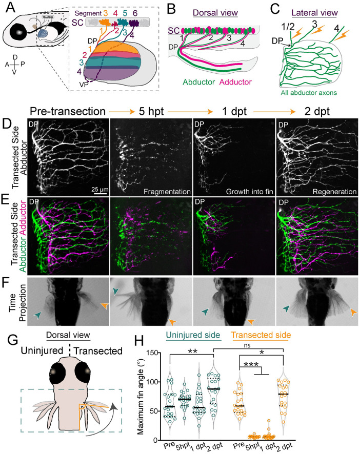
Fig 1 Pectoral fin motor axons regenerate robustly and reform functional synapses.

(A) Schematic of a 5 dpf larval zebrafish. Inset shows motor pools from SC segments 3–6 that form pectoral fin nerves 1–4 in the body wall. Axons sort at a DP or VP and innervate the musculature of the pectoral fin topographically. Innervation domains are labeled 1–4 and shown in corresponding colors. (B) Dorsal view. Motor neurons in SC segments innervate either the abductor or adductor muscle. (C) Lateral view. Schematic of abductor innervation of the pectoral fin. Nerves were transected using a laser in the locations shown with the lightning bolts. (D, E) Images from the transected side of maximum projections of fin motor innervation labeled with Tubb:dsRed. This example is from the same animal through regeneration. At 5 h hpt, axons have fragmented. At 1 dpt, axons have started to grow into the fin. At 2 dpt, axons have regenerated. (D) Abductor muscle innervation. (E) Abductor and adductor innervation pseudo-colored in green and magenta, respectively. (F) Corresponding time projections (<700 ms) of spontaneous pectoral fin movements. The region shown is indicated by the green dotted box in G. Only the nerves of the right fin were transected. At 5 hpt, the transected fin does not move. At 1 dpt, axons have just begun to grow into the fin but the transected fin still does not move. However, by 2 dpt, axons have regenerated and the transected fin can move again. The green and orange arrows point to the maximum fin position for the uninjured and transected sides, respectively. (G) The maximum angle of the tip of the fin compared to the body wall was measured during spontaneous fin movements. (H) Quantification of the maximum angle of the uninjured and transected fins pre-transection through regeneration. Each dot represents 1 fish and 1 movement. The black bar represents the mean. One-way ANOVA. *p = 0.04, **p < 0.005, ***p < 0.0001, ns = not significant. Original data for panel H is in S1 Data. dpf, day post fertilization; dpt, day post transection; DP, dorsal plexus; hpt, hour post transection; SC, spinal cord; VP, ventral plexus.

Acknowledgments
This image is the copyrighted work of the attributed author or publisher, and ZFIN has permission only to display this image to its users. Additional permissions should be obtained from the applicable author or publisher of the image. Full text @ PLoS Biol.