
Image
|
Figure Caption
Fig. 8
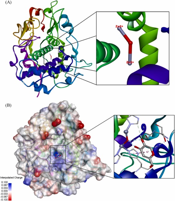
Fig. 8. Protein structure of TYRP-1 (5M8M) obtained from protein databank. (A) Image of the binuclear zinc active site for protein binding. (B) The binding pocket of ligands is colour coded with an interpolated charged surface (blue/red = positive/ negative).(For interpretation of the references to colour in this figure legend, the reader is referred to the web version of this article.)
Acknowledgments
This image is the copyrighted work of the attributed author or publisher, and
ZFIN has permission only to display this image to its users.
Additional permissions should be obtained from the applicable author or publisher of the image.
Full text @ Biomed. Pharmacother.