
|
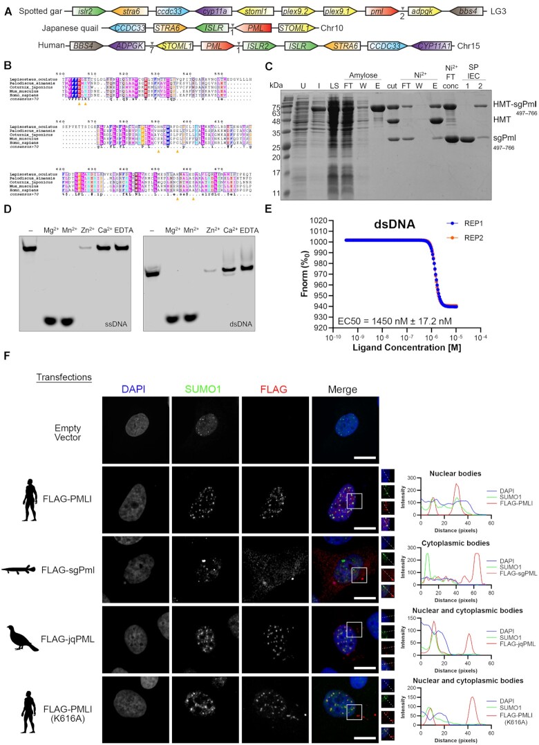
Fig. 2
Spotted gar Pml is an active cytoplasmic DNA exonuclease. (A) Synteny of pml locus between the genomes of the Spotted gar, Japanese quail and Humans. Similarly coloured genes represent homologs between species. The intervening arrows with numbers indicate the number of adjacent genes between homologous genes. (B) MUSCLE alignment of the spotted gar PML (sgPml) C-terminal DEDDh exonuclease (CDE) domain protein with other PML orthologs. The consensus sequence is displayed below the sequences for residues with over 70% conservation across species. Yellow arrows indicate predicated catalytic residues for sgPml and the red circle indicates the conserved SUMOylation site (PML-I K616). (C) Purification of sgPml-CDE. sgPml-CDE was expressed as a fusion to sequences encoding for hexahistidine, maltose binding protein, and a TEV protease cleavage site (HMT). SDS-PAGE analysis is shown for samples of uninduced (U) and induced (I) cells, soluble lysate (LS), after TEV cleavage (cut), flowthrough (FT), wash (W), and elution (E) fractions of amylose and Ni2+ affinity chromatography, and fractions from ion exchange chromatography (IEC). (D) Exonuclease activity of sgPml-CDE requires Mg2+ or Mn2+. Fluorescent oligonucleotides were incubated with sgPml-CDE in the presence of the indicated divalent cation or EDTA. ‘-’ refers to input without the addition of cations. (E) sgPml binds to dsDNA with high affinity. Microscale thermophoresis was used to quantify the affinity between sgPml-CDE and dsDNA (n = 2). The different coloured lines are data shown for two replicates. (F) Subcellular localizations of FLAG-tagged PML-I, sgPml, quail (jqPML) and PML-I (K616A). Cells were transfected with either an empty vector or the indicated FLAG-tagged proteins. Colocalization between FLAG-tagged proteins and SUMO1 was assessed by immunostaining and shown at the right as line plot for the region of interest bound by the white box in the merged fluorescence image. Additional fields of view are shown in Supplementary Figure S16. Scale bars represent 10 μm.