Fig. 5
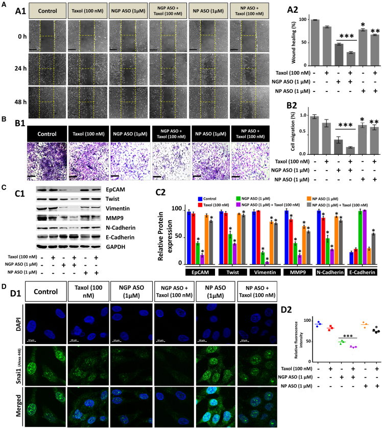
NGP ASO and Taxol synergistically prevent EMT in MCF7 cells
(A) Wound-healing assay where the width of wound closure in MCF7 cells at 0 h was set to 100% (A1). (A2) Percentage of wound closure plotted graphically with respect to control. Wound closure was most inhibited in the NGP ASO + Taxol group. (B) Transwell migration assay in MCF7 cells after combined compound treatment at indicated doses (B1). The number of cells that invaded to the lower chamber through Matrigel in the control sample is taken as 100%. (B2) Bar diagram shows the percentage of Transwell migration in MCF-7 cells of treated groups with respect to untreated control. Scale bars, 60 μm. (C) Western blot analysis for expression profiling of EpCAM, Twist, vimentin, MMP9, N-cadherin, and E-cadherin (C1) with their densitometric data (C2). (D) Confocal immunofluorescence microscopic analysis of Snai1 (shown in green) in control and treated cells after 72 h. Nuclei were stained with DAPI (blue). Quantification of Snai1 intensity per nucleus was obtained from confocal immunofluorescence microscopy and was calculated for 20–25 cells. Data presented as mean ± SEM (n = 3). ∗p < 0.05.

