
Image
|
Figure Caption
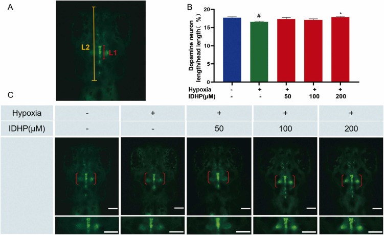
Fig. 2 IDHP protected the development of dopaminergic neurons (DANs) from hypoxia. (A) The schematic picture of dopaminergic neurons (DANs) (L1) and head (L2) measurement. (B) Statistical analysis of the ratio of L1/L2. The scale bar is 100 µm. n = 8. (C) Typical fluorescent images of DAN in Tg(vmat2:GFP) zebrafish larvae. Dopaminergic neuron (DAN)-rich region was delineated with red brackets. #P < 0.05, vs control group; *P < 0.05, vs hypoxia-treated group.
Acknowledgments
This image is the copyrighted work of the attributed author or publisher, and
ZFIN has permission only to display this image to its users.
Additional permissions should be obtained from the applicable author or publisher of the image.
Full text @ Biomed. Pharmacother.