
Fig. 2
- ID
- ZDB-IMAGE-230828-96
- Publication
- Domingues et al., 2023 - Cholesteryl hemiazelate Identified in Cardiovascular Disease Patients Causes in vitro and in vivo Inflammation
- All Figures
- Figures for Domingues et al., 2023
|
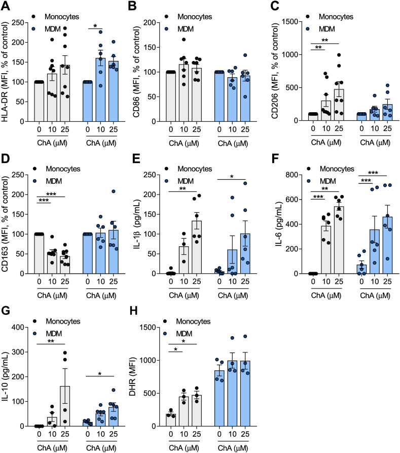
Fig. 2
ChA changes the expression of surface markers and the inflammatory profile of human primary monocytes (Mono) and MDMs. Peripheral blood was harvested from human volunteers. Mononuclear leukocytes (peripheral blood mononuclear cells) were isolated from whole blood using histopaque-1077. CD14+ cells were purified from total peripheral blood mononuclear cells by immunomagnetic separation. CD14+ monocytes were cultured in the presence of ChA (10 μM or 25 μM) or vehicle for 24 h (eight different donors) or for 7 days to obtain MDM (six different donors). Monocytes were differentiated using rM-CSF. After lipid treatment, a representative sample of cells was stained with monoclonal antibodies to determine surface expression of HLA-DR (A), CD86 (B), CD206 (C), and CD163 (D); data represent the mean fluorescence intensity (MFI) ± SEM for all subjects normalized to the control cells. Cell culture supernatants were analyzed by ELISA for IL-1β (E), IL-6 (F), and IL-10 release (G). Values represent the mean ± SEM of cytokine released (in pg/ml). Reactive oxygen species production by monocytes and MDM exposed to ChA (H). ∗