
Image
|
Figure Caption
Fig. 3
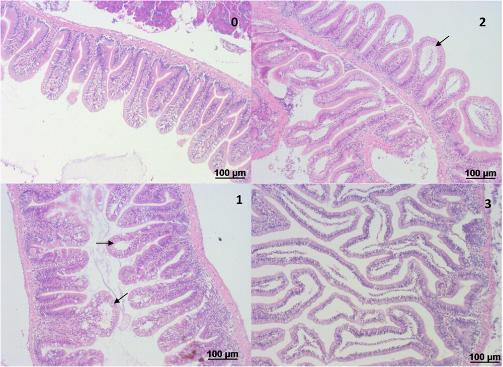
Hindgut of adult zebrafish following challenge with 120 mM TNBS, applied by anal intubation. The gut was analyzed 24 h after TNBS intubation for pathological alterations. The most prominent and consistent alteration was epithelial detachment (shown with arrow), which was graded to the degree of severity. The grade is noted on the top right of each image as follows: Level 0: gut of naïve zebrafish; Level 1: epithelial detachment is restricted to the top of the villus and accounts for 15–20% of the epithelium; Level 2: epithelial detachment involves about half of the villi and accounts for 50% of the epithelium; Level 3: epithelial detachment involves the whole length of the villus and accounts for 90–95% of the epithelium. H&E staining.
Acknowledgments
This image is the copyrighted work of the attributed author or publisher, and
ZFIN has permission only to display this image to its users.
Additional permissions should be obtained from the applicable author or publisher of the image.
Full text @ Mol. Nutr. Food Res.