
Image
|
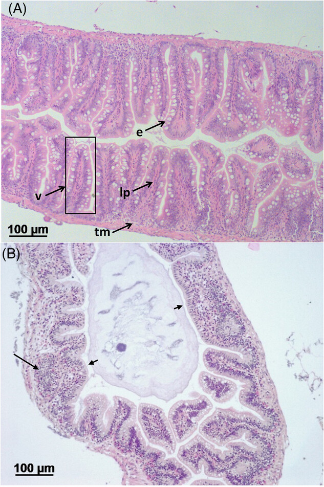
Figure Caption
Fig. 2
Administration of TNBS resulted in pathological alterations in the intestine. A) Normal structure of intact zebrafish intestine. V, villus; e, epithelium with goblet cells; lp, lamina propria; tm, tunica muscularis. B) Gut tissue histopathology in TNBS-challenged zebrafish at 24 h post-TNBS intubation. Long arrows point at infiltration of leucocytes into intestinal villi; short arrows point at enlarged villus. H&E staining.
Acknowledgments
This image is the copyrighted work of the attributed author or publisher, and
ZFIN has permission only to display this image to its users.
Additional permissions should be obtained from the applicable author or publisher of the image.
Full text @ Mol. Nutr. Food Res.