IMAGE

Fig. 6

ID
ZDB-IMAGE-230814-129
Source
Figures for Tang et al., 2021
Image
Figure Caption

Fig. 6

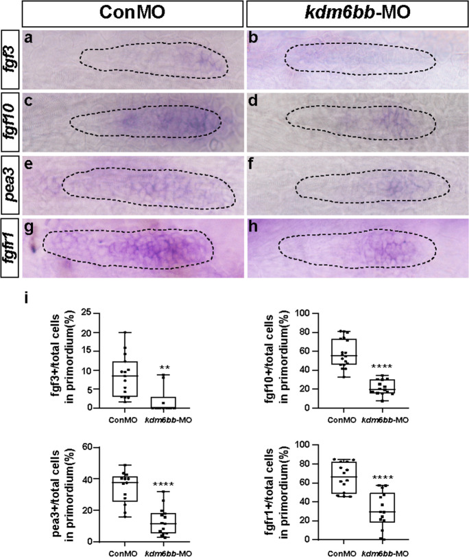
Kdm6b-depletion represses Fgf signaling in zebrafish primordium. a–h Corresponding decreased expression of Fgf signaling components including fgf3, fgf10, pea3, and fgfr1 are presented in kdm6bb morphants compared to the control siblings at 32 hpf. i The ratio of fgf3, fgf10, pea3, and fgfr1-positive cells to total cells in the primordia of controls (ConMO; n = 15 embryos for fgf3, n = 18 embryos for fgf10, n = 13 embryos for pea3, and n = 14 embryos for fgfr1) and experiment group (kdm6bb-MO; n = 10 embryos for fgf3, n = 14 embryos for fgf10, n = 14 embryos for pea3, and n = 14 embryos for fgfr1).The primordium appearance is shaped by dotted lines. Data are recorded as mean (minimum and maximum values). **p < 0.01, and ****p < 0.0001

Acknowledgments
This image is the copyrighted work of the attributed author or publisher, and ZFIN has permission only to display this image to its users. Additional permissions should be obtained from the applicable author or publisher of the image. Full text @ Cell Biol. Toxicol.