IMAGE

Fig. 2

ID
ZDB-IMAGE-230814-124
Source
Figures for Tang et al., 2021
Image
Figure Caption

Fig. 2

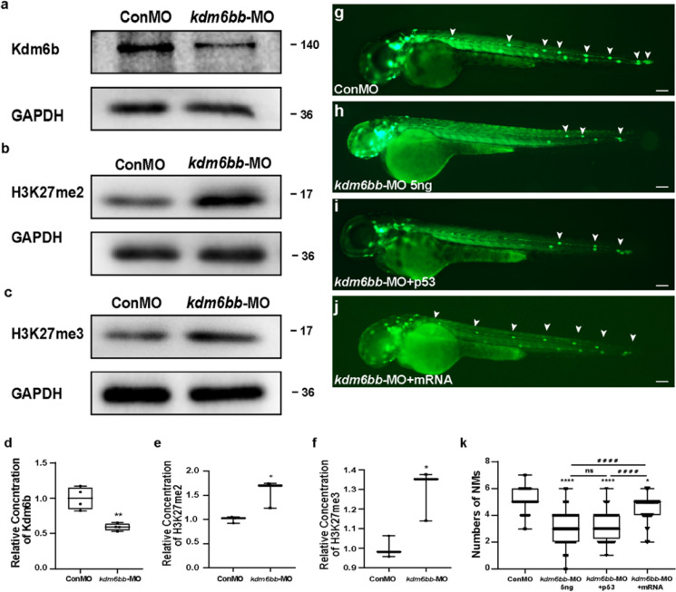
Kdm6b is required for cell migration and neuromast deposition in zebrafish posterior lateral lines. a–f Kdm6b level is effectively downregulated using the morpholino technology. The protein blotting of Kdm6b is significantly decreased (a), and the protein blottings of H3K27me2 (b) and H3k27me3 (c) were both upregulated by the special antisense morpholino injection both in the band intensity. d–f The quantification analysis of relative concentration of Kdm6b, H3K27me2, and H3K27me3. Data are recorded as mean (minimum and maximum values). *p < 0.05 and **p < 0.01. g–j At 48 hpf, the deposited neuromasts are labeled in green along the posterior body of control embryos (g), Kdm6b-deficient mutants (h), co-injection of kdm6bb-MO + p53 (i), and co-injection of kdm6bb-MO + kdm6bb mRNA specimens (j). The neuromasts of PLL are labeled in green fluorescence in the transgenic cldnb:lynGFP embryos, and a severe reduction in number of neuromasts is found (g–h). The decreased number of neuromasts was confirmed when co-injected with p53 and kdm6bb-MO (i). The decreased number of neuromasts by Kdm6b-defect is partially rescued by the combined injection of kdm6bb mRNA (j). White arrowheads label the neuromast along the trunk and terminal of the posterior LL (g–j). k Statistical analysis of the number of posterior LL neuromasts at 48 hpf in controls (n = 185), Kdm6b-deficient embryos (n = 129), and kdm6bb-MO + mRNA members (n = 42). ****p < 0.0001 (the contrast between kdm6bb-MO group with Con-MO group), ####p < 0.0001 (the contrast between kdm6bb-MO group with kdm6bb-MO + mRNA group), *p < 0.05 (the contrast between kdm6bb-MO + mRNA group with Con-MO group). Scale bars mark a distance of 100 μm

Acknowledgments
This image is the copyrighted work of the attributed author or publisher, and ZFIN has permission only to display this image to its users. Additional permissions should be obtained from the applicable author or publisher of the image. Full text @ Cell Biol. Toxicol.