
|
Fig. 5
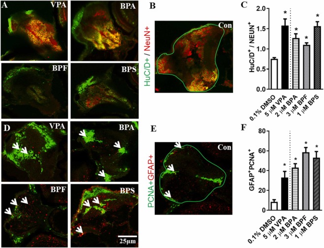
Fig. 5. BPA, BPF, and BPS accelerate neurogenesis. Zebrafish embryos exposed to 0.1 %DMSO (vehicle control), BPA (2 μM), BPF (3 μM), BPS (1 μM), and VPA (5 μM; positive control) from 8 to 108 hpf were assessed for neurogenesis at 4.5 dpf. (A) Staining patterns of mature newborn neurons (HuC/D+; green) and mature neurons (NeuN+; red) on tissue sections from the anterior telencephalon position. (B) The region of interest in the anterior telencephalon used for fluorescence intensity quantification. (C) The ratio of fluorescence intensity of HuC/D+ to NEUN+ (N = 6–10). (D) Staining patterns of neural stem cells (GFAP+; red) and cell proliferation marker (PCNA+; green) on tissue sections from the anterior telencephalon position. (E) The region of interest in the anterior telencephalon used for counting GFAP+PCNA+ cells. Arrows indicate double positive cells. (F) Quantification of GFAP+PCNA+ cells (N = 4–10). Values are plotted as the mean ± SEM, and the asterisk (*) indicates a significant difference from the vehicle control (0.1 % DMSO) at P < 0.05. Scale bar, 25 µm.