
|
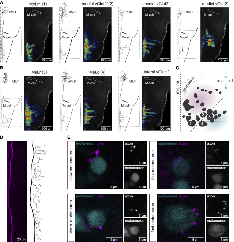
Fig. 6 Pattern of projection of nMLF neurons
(A) Morphologies and collateral projections in hindbrain of four medial vGlut2+ nMLF neurons. For each neuron type, the left panel shows reconstruction of a single neuron and the right panel shows heatmap of the collaterals of all recorded neurons.
(B) Morphologies and collateral projections in hindbrain of three lateral vGlut1+ nMLF neurons. For each neuron type, the left panel shows reconstruction of a single neuron and the right panel shows heatmap of the collaterals of all recorded neurons.
(C) Location of the analyzed neurons in the nMLF showing the soma position of four large neurons (1, MeLm; 2, large medial neuron; 3, MeLr; 4, MeLc).
(D) Confocal image and reconstruction of the axonal projection of an nMLF neuron in the rostral spinal cord.
(E) Close contacts between axon collaterals of nMLF neurons and motoneurons of the slow, intermediate, and fast modules.