
|
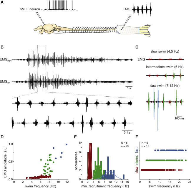
Fig. 3 Spontaneous swim activity in the absence of sensory input in adult zebrafish
(A) Experimental setup for ex vivo recordings of nMLF neurons during spontaneous swimming activity.
(B) Representative example of a spontaneous swimming episode recorded as EMG activity on the right and left body sides. Enlarged portion shows coordinated swim pattern with alternating muscle activity between the right and left body sides.
(C) The amplitude of the EMG activity increased with increasing swim frequency. Small slow muscle units (red) were recruited during slow swimming (top, 4.5 Hz swim frequency). Both slow (red) and intermediate (green) muscle units were recruited during intermediate swim (middle, 6 Hz swim frequency). Slow (red), intermediate (green), and large fast (blue) muscle units were all recruited during fast swimming (bottom, 7–12 Hz swim frequency).
(D) Recruitment of slow (red), intermediate (green), and fast (blue) muscle units with increasing swim frequency. Each dot denotes one swim cycle pooled from several swim episodes in a single animal.
(E) Minimum recruitment frequencies of the slow (red), intermediate (green), and fast (blue) units observed in 29 swim episodes in 5 fish.
(F) Activity span of different muscle units (slow, red; intermediate, green; fast, blue). Each dot denotes one swim cycle during which the respective muscle units were active (data from n = 15 swim episodes in N = 5 fish).