
Image
|
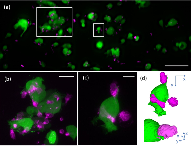
Figure Caption
Fig. 7
Hybrid tiled projection and targeted 3D imaging. (a) Last frame of series of tiled projection images of NK cells labeled with CellTracker red (shown in magenta) and MCF7 breast cancer cells cytosolically labeled with GFP (shown in green). (b) Projection of a 3D stack acquired around the large white rectangle in (a). (c) Projection of a 3D volume acquired around the small white rectangle in (a). (d) Rendering of the cell shown in (c). Scale bars: (a) 100 µm, (b) 30 µm, (c) 10µm.
Acknowledgments
This image is the copyrighted work of the attributed author or publisher, and
ZFIN has permission only to display this image to its users.
Additional permissions should be obtained from the applicable author or publisher of the image.
Full text @ Biomed. Opt. Express