Image
Figure Caption
Fig. 3
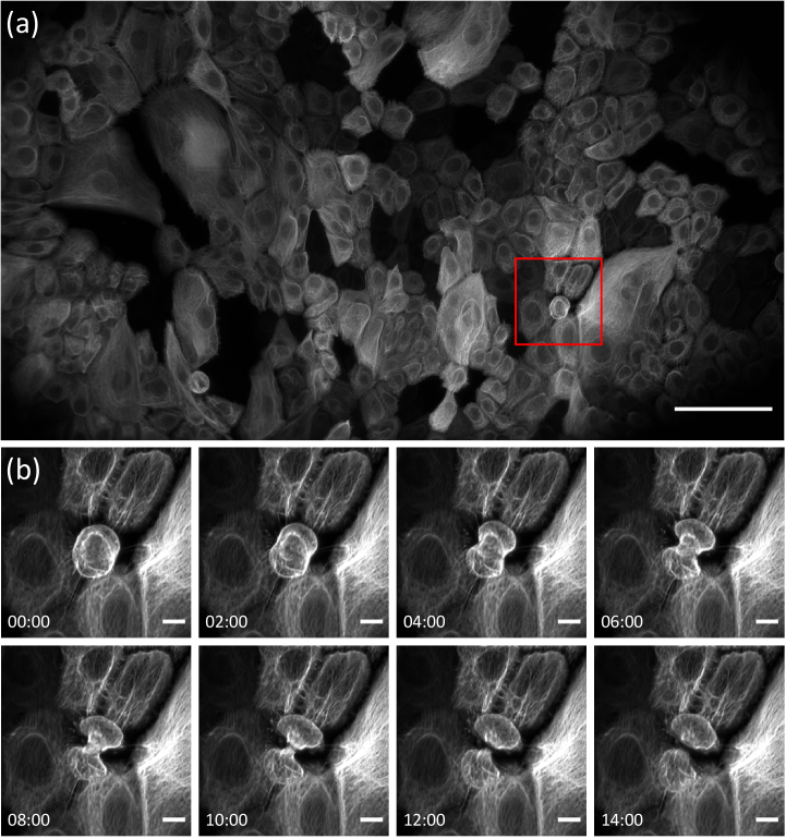
Imaging of the intermediate filament cytoskeleton in a monolayer of cultured keratinocytes expressing keratin 5 (K5)-mNeonGreen. (a) Maximum intensity projection of the tiled volume. A gamma correction of 0.7 has been applied. (b) Zoom on the red boxed region in (a), demonstrating profound rearrangement of the intermediate filament network during cell division. Scale bars: (a) 100 µm, (b) 10 µm. Timestamp: mm: ss.
Acknowledgments
This image is the copyrighted work of the attributed author or publisher, and
ZFIN has permission only to display this image to its users.
Additional permissions should be obtained from the applicable author or publisher of the image.
Full text @ Biomed. Opt. Express

