
Fig. 5
- ID
- ZDB-IMAGE-230703-29
- Genes
- Publication
- Fetsko et al., 2022 - Brain endothelial cells acquire blood-brain barrier properties in the absence of Vegf-dependent CNS angiogenesis
- All Figures
- Figures for Fetsko et al., 2022
|
Fig. 5
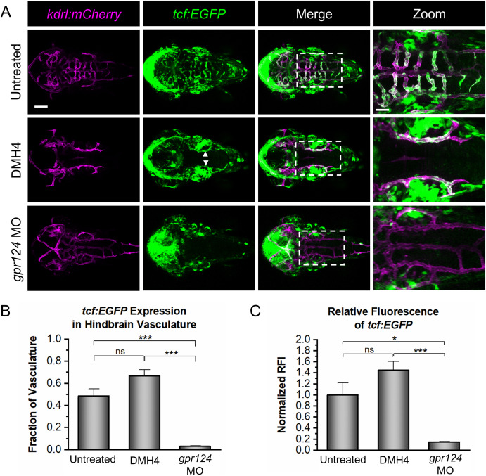
Fig. 5. Vegf signaling is not required for Wnt/β-catenin transcriptional activity. (A) Representative confocal images of tcf:EGFP, kdrl:mCherry embryos at 2 dpf that were untreated, DMH4-treated (DMH4), or injected with a gpr124 morpholino (gpr124 MO). tcf:EGFP signal is present in the WT brain vasculature (top panels) and in the PHBCs of the DMH4-treated embryos (middle panels; white arrows), but not the gpr124 MO-injected embryos (bottom panels). Scale bar is 100 μm for the first three columns and 40 μm for the zoomed images (right panels). (B) Quantification of the fraction of kdrl:mCherry-labeled hindbrain vasculature expressing tcf:EGFP. (C) Quantification of the normalized relative fluorescence intensity (RFI) of tcf:EGFP in the hindbrain vasculature. Data in B and C are presented as means (n = 4) ± SEM (∗p < 0.05; ∗∗∗p < 0.001; ns = not significant).
Reprinted from Developmental Biology, 494, Fetsko, A.R., Sebo, D.J., Taylor, M.R., Brain endothelial cells acquire blood-brain barrier properties in the absence of Vegf-dependent CNS angiogenesis, 46-59, Copyright (2022) with permission from Elsevier. Full text @ Dev. Biol.