
Fig. 4
- ID
- ZDB-IMAGE-230703-28
- Antibodies
- Publication
- Fetsko et al., 2022 - Brain endothelial cells acquire blood-brain barrier properties in the absence of Vegf-dependent CNS angiogenesis
- All Figures
- Figures for Fetsko et al., 2022
|
Fig. 4
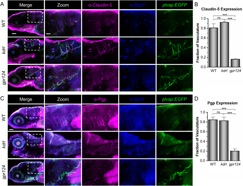
Fig. 4. Zebrafish kdrl, but not gpr124, mutants, express BBB markers. (A) Representative confocal microscopy images showing Claudin-5 staining. WT, kdrl, and gpr124 larvae were stained with ⍺-Claudin-5 and ⍺-Glut1. The plvap:EGFP transgene was used as a blood vessel marker. (B) Quantification was calculated from the fraction of the total blood vessels in the hindbrain labeled with ⍺-Claudin-5. (C, D) Representative confocal images of ⍺-Pgp staining at 5 dpf with the same controls as in (A) and quantification strategy as in (B). All images are lateral views (anterior left; dorsal top) of whole-mount stained larvae at 5 dpf. Scale bars are 100 μm for the merged images (left panels) and 40 μm for the zoomed images. Data in B and D are presented as means (n = 3) ± SEM (∗∗∗p < 0.001; ns = not significant).
Reprinted from Developmental Biology, 494, Fetsko, A.R., Sebo, D.J., Taylor, M.R., Brain endothelial cells acquire blood-brain barrier properties in the absence of Vegf-dependent CNS angiogenesis, 46-59, Copyright (2022) with permission from Elsevier. Full text @ Dev. Biol.