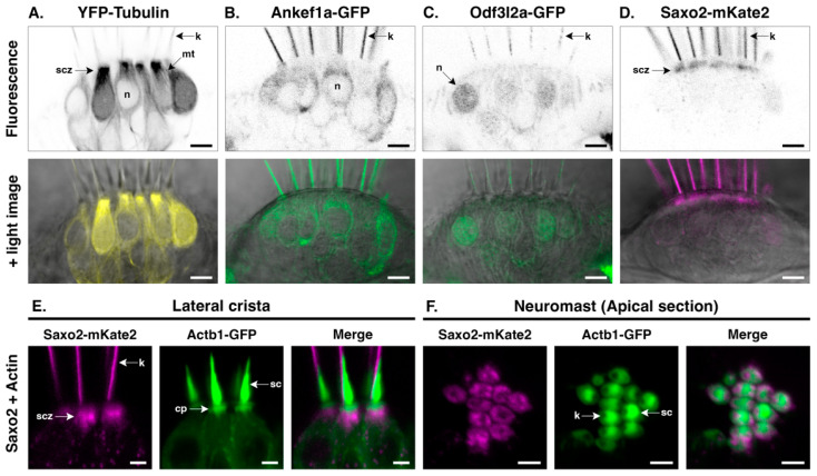
Distinct protein localization patterns in the hair cell soma. (A–D) Details of YFP-TUBA, Ankef1a-GFP, Odf3l2a-GFP, and Saxo2-mKate2 in the hair cell body at 5–6 dpf. The top panels are black-and-white representations of the fluorescence channel and the lower panels overlay the fluorescence channel with the corresponding light image. (E,F) Saxo2-mKate2 (magenta) accumulations in the apical part of the hair cell body in cristae and neuromasts. GFP-tagged beta-actin is shown in green. cp = cuticular plate; k = kinocilium; mt = microtubule; n = nucleus; sc = stereocilia; scz = subcuticular zone. Scale bars are 5 µm in (A–D) and 2.5 µm in (E,F).
Acknowledgments
This image is the copyrighted work of the attributed author or publisher, and
ZFIN has permission only to display this image to its users.
Additional permissions should be obtained from the applicable author or publisher of the image.
Full text @ J Dev Biol
Your Input Welcome
Thank you for submitting comments. Your input has been emailed to ZFIN curators who may contact you if
additional information is required.
Oops. Something went wrong. Please try again later.