IMAGE

Figure 4

ID
ZDB-IMAGE-230621-48
Source
Figures for Ochi et al., 2020
Image
Figure Caption

Figure 4

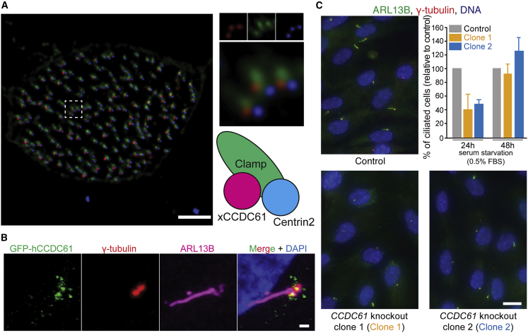
CCDC61 Associates with Basal Bodies and Plays a Role in Ciliogenesis

(A) xCCDC61 associates with basal bodies and rootlets in multi-ciliated epidermal cells of Xenopus embryos. A fluorescent image of a Xenopus embryo expressing xCCDC61-RFP (red), the basal body component Centrin2-BFP (blue), and the rootlet component Clamp-GFP (green). Scale bar, 3 μm.

(B) Location of hCCDC61 at the periphery of basal bodies of primary cilia. Immunofluorescent image of an RPE-1 cell transiently overexpressing GFP-hCCDC61. Co-immunofluorescent staining was performed against GFP (green), basal bodies (γ-tubulin, red), and the ciliary axoneme (ARL13B, magenta). Scale bar, 1 μm.

(C) Ciliated cells of control and CCDC61-knockout RPE-1 cells. Immunofluorescent images show representative immunofluorescent images used for quantifications of ciliogenesis of primary cilia. Scale bar, 10 μm. The bar graph shows that ciliogenesis was delayed in the CCDC61 knockout cells. Data shown correspond to three biological replicates (total cell counts n = 1,181, 1,103, and 1,008 for control, clone 1 and clone 2 cells after 24-h serum starvation respectively, and n = 1,151, 1,046 and 1,242 for control, clone 1 and clone 2 after 48-h serum starvation, respectively). Percentages are relative to control cells. Bar graphs show mean ± standard deviation.

See also Figure S5.

Acknowledgments
This image is the copyrighted work of the attributed author or publisher, and ZFIN has permission only to display this image to its users. Additional permissions should be obtained from the applicable author or publisher of the image.

Reprinted from Structure (London, England : 1993), 28, Ochi, T., Quarantotti, V., Lin, H., Jullien, J., Rosa E Silva, I., Boselli, F., Barnabas, D.D., Johnson, C.M., McLaughlin, S.H., Freund, S.M.V., Blackford, A.N., Kimata, Y., Goldstein, R.E., Jackson, S.P., Blundell, T.L., Dutcher, S.K., Gergely, F., van Breugel, M., CCDC61/VFL3 Is a Paralog of SAS6 and Promotes Ciliary Functions, 674689.e11674-689.e11, Copyright (2020) with permission from Elsevier. Full text @ Structure