
Image
|
Figure Caption
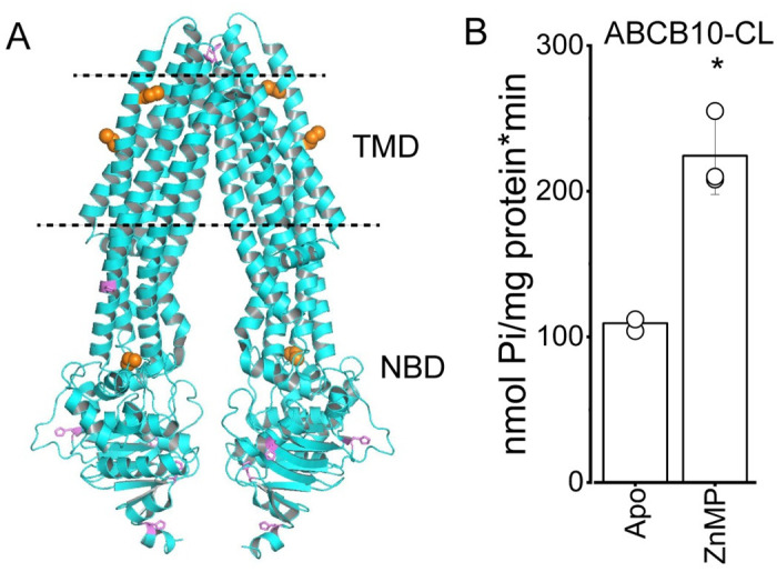
Fig 4 Cysteine residues in ABCB10 are not necessary for ZnMP activation
(A) Structural model of the ABCB10 homodimer (PDB 3ZDQ, both monomers in cyan), with histidine residues shown as violet sticks and cysteine residues as orange spheres. The expected phospholipid membrane borders are indicated by the dashed lines. TMD and NBD correspond to the transmembrane domain and nucleotide binding domain, respectively. Figure was created in PyMOL. (B) ATPase activity of cysteine less ABCB10 (ABCB10-CL) in the absence (apo) and presence of 5 μM ZnMP (n = 3; p = 0.05).
Acknowledgments
This image is the copyrighted work of the attributed author or publisher, and
ZFIN has permission only to display this image to its users.
Additional permissions should be obtained from the applicable author or publisher of the image.
Full text @ PLoS One