IMAGE

Fig 11

ID
ZDB-IMAGE-230621-20
Source
Figures for Guan et al., 2016
Image
Figure Caption

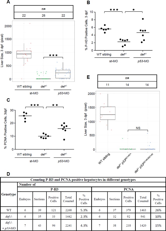
Fig 11 Def regulates liver development only in part through the p53 pathway.

(A–D) WISH analysis of liver development (A) and histogram showing the statistical data for the ratio of P-H3- (B) or PCNA- (C) positive cells in def-/- embryos injected with p53-specific morpholino mixes (ATG-MO plus spl-MO) at 3 dpf. Number of embryos used for sectioning and number of sections used for counting P-H3- and PCNA-positive hepatocytes in different genotypes in (B) and (C) was shown in (D). (E) WISH analysis of liver development in the def-/-p53M214K double mutant at 3dpf. The fabp10a probe was used for WISH. Liver size is measured by liver area marked by the fabp10a-positive signal. In the quartile boxplot, each dot represents the liver size of an individual embryo. st-MO, standard control morpholino; ATG-MO, morpholino targeting the translation start site (ATG) of p53; spl-MO, morpholino targeting the splicing junction of exon 5 and intron 5 of p53. In (A) and (E), total number of embryos used for each genotype is shown on the top of the graph. Underlying data for (A), (B), (C), and (E) are provided in S1 Data.

Acknowledgments
This image is the copyrighted work of the attributed author or publisher, and ZFIN has permission only to display this image to its users. Additional permissions should be obtained from the applicable author or publisher of the image. Full text @ PLoS Biol.