IMAGE

Fig 1

ID
ZDB-IMAGE-230621-10
Source
Figures for Guan et al., 2016
Image
Figure Caption

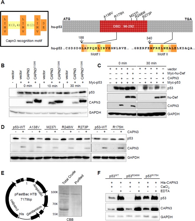
Fig 1 Wild-type p53, but not the mutant p53R175H, is a substrate of CAPN3.

(A) Left panel: the CAPN3 recognition motif. Right panel: upper panel shows the simplified hu-p53 protein with five frequent mutated sites in the DNA-binding domain (DBD) highlighted. Lower panels depict the two putative CAPN3 recognition regions in p53. Numbers denote the position of amino acids in p53. ATG, start codon; TGA, stop codon. (B–D) In vitro assay of p53 degradation by CAPN3 or CAPN3C129S. CAPN3, CAPN3C129S, Myc-hu-Def, Myc-p53, and its mutant derivatives were expressed in 293T cells, respectively. Protein extracts were mixed as indicated and the mixture was incubated in the reaction buffer containing 5 mM CaCl2 at 37°C for 0, 10, 30 min (B), or 30 min (C), or 45 min (D). GAPDH: loading control. Immunodetection of p53 was achieved by using a combination of monoclonal antibodies PAb240 and PAb1620 (B, D), or Anti-Myc tag antibody 9E10 (C). GAPDH was recognised by mouse monoclonal antibody 5-E10 (B) or rabbit monoclonal antibody EPR1977Y (C, D). (E) Left panel: diagram showing the CAPN3-pFastBac plasmid (baculovirus expression system) for transfecting the SF9 cells to express His-CAPN3. Right panel: Coomassie brilliant blue (CBB) staining picture showing total protein crude extract (total crude) and purified His-CAPN3 using the Ni-NTA agarose beads (purified). (F) In vitro assay of the degradation of Myc-p53WT-HA, Myc-p53R248W-HA, or Myc-p53R175H-HA by the purified His-CAPN3 in the presence of 10 mM CaCl2 or 20 mM EDTA as indicated at 37°C for 45 min. GAPDH: loading control. p53 was detected by a combination of monoclonal antibodies PAb240 and Pab1620. GAPDH was recognised by mouse monoclonal antibody 5-E10.

Acknowledgments
This image is the copyrighted work of the attributed author or publisher, and ZFIN has permission only to display this image to its users. Additional permissions should be obtained from the applicable author or publisher of the image. Full text @ PLoS Biol.