IMAGE

Fig. 5

ID
ZDB-IMAGE-230619-16
Source
Figures for Groenewoud et al., 2023
Image
Figure Caption

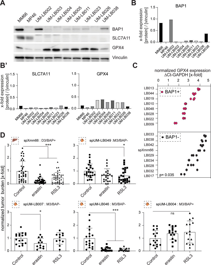
Fig. 5 Ferroptosis induction inhibits metastasis formation in the metastatic uveal melanoma zebrafish model.

A Western blot analysis determining GPX4, SLC7A11 and BAP1 protein expression in the two established cell lines MM66 and MM 46 and eight patient-derived tissues. Loading control: vinculin. Note, while BAP1 and GPX4 show a negative correlation in the patient samples, SLC7A11 was barely detected, independent of BAP1 expression levels. B, B’ Westernblot quantification of BAP1, SLC7A11 and GPX4. C qPCR analysis of GPX4 mRNA expression in primary UM tissues and spXmm66 (green dots), with known BAP1 status. Expression values were normalized to GAPDH (ΔCT). n = 3. D Quantitative analysis of tumor burden in the here established metastatic uveal melanoma zebrafish model. The indicated cells were engrafted at 48 hpf, maintained for 6 days in the absence or presence of the ferroptosis inducer erastin (SLC7A11 inhibitor, 5 μM) or RSL3 (GPX4 inhibitor, 10 μM) and subsequently subjected to tumor burden analysis. Notably, ferroptosis induction via erastin and/or RSL3 resulted in 4 out of 5 cases in a significant reduction of tumor burden. n = 40 (spXmm66) or n = 20 (all other cases). Data are mean ± SD. D3: chromosome 3 disomy: M3: chromosome 3 monosomy. ns: not significant. ***:p < 0.001.

Acknowledgments
This image is the copyrighted work of the attributed author or publisher, and ZFIN has permission only to display this image to its users. Additional permissions should be obtained from the applicable author or publisher of the image. Full text @ Cell Death Discov