IMAGE

Fig. 3

ID
ZDB-IMAGE-230619-14
Source
Figures for Groenewoud et al., 2023
Image
Figure Caption

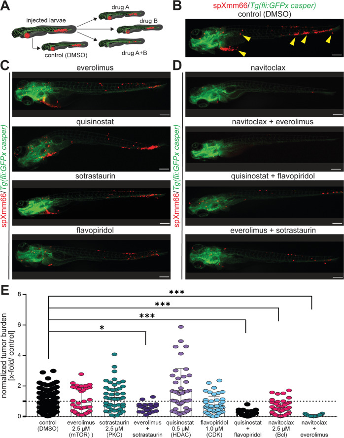
Fig. 3 Metastatic uveal melanoma zebrafish model is suitable for drug screening.

A Scheme representing the approach to perform drug screening in the here established metastatic uveal melanoma zebrafish model utilizing spXmm66 cell engraftment at 48 hpf followed by tumor burden analysis 6 dpi. BD Stereomicroscopic images of representative phenotypes of Tg(fliGFPx casper) zebrafish larvae 6 days post injection with tdTomato-labelled spXmm66 cells and treatment with the indicated drugs at their maximum tolerated dose (>80% survival, Supplementary Fig. 1). Scale bars: 250 µm. E Quantitative analysis of (BD) to determine the effect of the indicated drug treatment on tumor burden, normalized to DMSO control (normalized tumor burden). Data are mean ± SD. *:p < 0.05; **:p < 0.01; ***:p < 0.001.

Acknowledgments
This image is the copyrighted work of the attributed author or publisher, and ZFIN has permission only to display this image to its users. Additional permissions should be obtained from the applicable author or publisher of the image. Full text @ Cell Death Discov