IMAGE

Fig. 2

ID
ZDB-IMAGE-230619-12
Source
Figures for Groenewoud et al., 2023
Image
Figure Caption

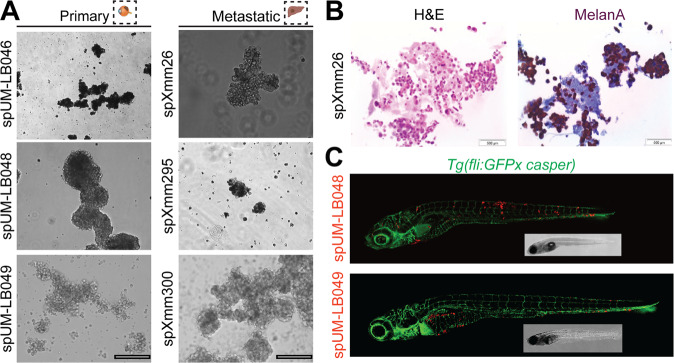
Fig. 2 Spheroid cultures can readily be established from both primary uveal melanoma tumors and patient-derived metastatic uveal melanoma tissues derived from murine xenografts.

A Representative image of the established spheroid cultures. Primary: uveal melanoma patient tumor tissue. Metastatic: murine xenograft material, derived from metastatic UM samples propagated subcutaneously. Scale bars: 250 µm). B Analysis of spXmm26 by H&E as well as melanA staining (marker for the melanocytic origin of the engrafted cells). C Stereomicroscopic images of representative phenotypes of Tg(fliGFPx casper) zebrafish larvae (blood vessels: green) 6 days post injection with CM-DiL-labelled spheroid cells (spUM-LB048 and spUM-LB049, red) derived from primary uveal melanoma tumor tissues. Note, disseminated cancer cells are present up to 6 days post engraftment and settle in both the hematopoietic tissue and/or the liver. Inserts: same fish imaged with bright field microscopy.

Acknowledgments
This image is the copyrighted work of the attributed author or publisher, and ZFIN has permission only to display this image to its users. Additional permissions should be obtained from the applicable author or publisher of the image. Full text @ Cell Death Discov