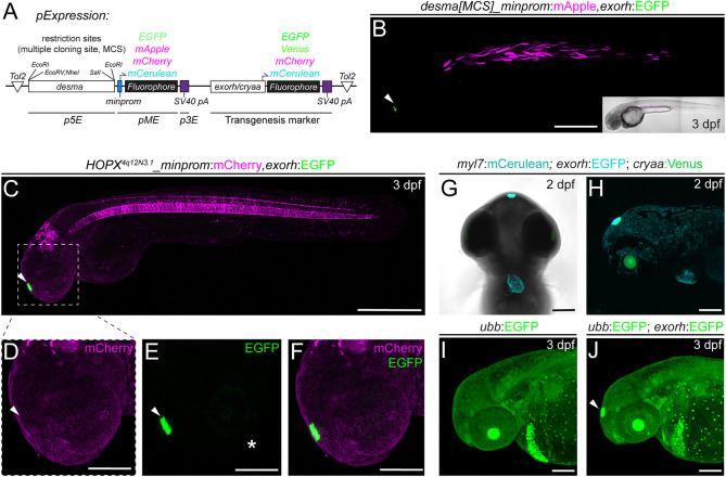
IMAGE

Fig. 6.

ID
ZDB-IMAGE-230617-40
Source
Figures for Kemmler et al., 2023
Image
Figure Caption

Fig. 6.

Application examples of an exorh-based, pineal gland-specific transgenesis reporter. (A,B) Injection-based, transient testing and stable transgenesis of regulatory elements with Tol2 vectors that harbor a transgenesis marker in cis for quality control. The desmin a (desma) upstream region paired with the mouse beta-globin minimal promoter driving different fluorophores with exorh- or cryaa-based (eye lens) transgenesis markers in cis in Tol2 backbones (A, see also Table 1). In all vector versions, the desma regulatory region is flanked by restriction sites known as simple Multiple Cloning Sites (MCSs), enabling restriction enzyme-based replacement of desma with a regulatory element of interest (A). (B) Injection shows muscle-selective desma activity (mCherry) and pineal-specific transgenesis reporter expression (EGFP). (C-F) Regulatory element testing with exorh:EGFP as a transgenesis marker in cis. Stable transgene integration of HOPX4q12N3.1:minprom-mCherry,exorh:EGFP at 3 dpf shows mCherry expression in the notochord and rhombomeres, whereas EGFP expression is confined to the pineal gland (C, arrowhead). The transgenes do not cross-activate mCherry (D,F) or EGFP (E,F) (insets, white arrowheads), despite being encoded in cis and in close proximity, suggesting the exorh regulatory region is inert. The white asterisk (E) indicates faint patchy EGFP signal in the eye. (G,H) Combinatorial use of transgenesis markers. Ventral (G) and lateral (H) views of a stable myl7:mCerulean,exorh:EGFP transgenic crossed to cryaa:Venus and imaged at 2 dpf; there are distinct expression patterns of the secondary transgenic markers labeling the heart (myl7), pineal gland (exorh) and eye lens (cryaa), respectively. (I,J) exorh:EGFP fluorescence is discernible in ubiquitous green-fluorescent transgenics. Ubiquitous ubb:EGFP (ubi:Switch) expression (I) still allows detection of exorh:EGFP expression (white arrowhead) (J). Scale bars: 500 μm (B,C); 200 μm (D-J).

Acknowledgments
This image is the copyrighted work of the attributed author or publisher, and ZFIN has permission only to display this image to its users. Additional permissions should be obtained from the applicable author or publisher of the image. Full text @ Development