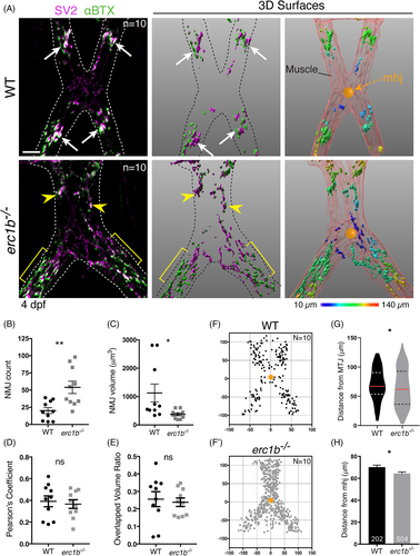
Fig. 5 Small, diffusely distributed NMJs appear in Erc1b deficient larvae. A, Left: IF labeling with SV2 (magenta) and αBTX (green); dotted line outlines jaw muscles. Large clusters of NMJs (white arrows) form in WT, erc1b−/− larvae clusters (yellow arrowheads) disperse throughout the tissue (yellow brackets). Middle: Imaris 3D renderings of NMJs. Right: Pseudo-colored NMJs based on the distance from mandibulohyoid junction (mhj, orange ball) (see Section 4 for details). B, Quantification of the number of NMJs, C, NMJ volume, D, SV2/αBTX colocalization Pearson's coefficient, and E, 3D overlap volume ratio in WT and erc1b−/− larvae. Symbols indicate individual embryos (WT N = 10, erc1b−/− N = 10), lines indicate mean with SEM. The location of each NMJ plotted in XY in reference to the mhj (0,0; orange ball) in WT, F, and erc1b−/−, F′, larvae. G, Violin plots of NMJ distance from the mhj (median = red line, upper and lower quartiles = dotted lines). H, NMJ distance to the mhj; bars indicate mean with SEM, total number of NMJs noted at the bottom of each bar (WT n = 202, erc1b−/− n = 504). Mann–Whitney U test (two-tailed) was used for statistical analysis, 95% confidence interval, *P < 0.05, **P < 0.01. All images are maximum intensity projections, ventral views with anterior on top. Scale bars = 25 μm
Image
Figure Caption
Figure Data
Acknowledgments
This image is the copyrighted work of the attributed author or publisher, and
ZFIN has permission only to display this image to its users.
Additional permissions should be obtained from the applicable author or publisher of the image.
Full text @ Dev. Dyn.

