IMAGE

Figure 3

ID
ZDB-IMAGE-230531-27
Source
Figures for Zhao et al., 2023
Image
Figure Caption

Figure 3

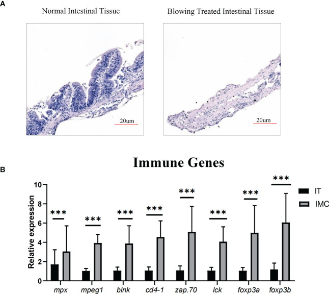
Enriching intestinal immune cells via stripping the muscular layer. (A) Morphological analysis of HE staining in comparison between intestinal tissue after blowing treatment and normal intestinal tissue. (B) qPCR analysis of genes involved in immune cell differentiation. All values are means ± SEM; statistical significance was determined by independent-samples T-test. For the comparison of IMC (intestinal mucosa cells) vs IT (intestinal tissue), *** represented p < 0.01.

Acknowledgments
This image is the copyrighted work of the attributed author or publisher, and ZFIN has permission only to display this image to its users. Additional permissions should be obtained from the applicable author or publisher of the image. Full text @ Front Immunol