
|
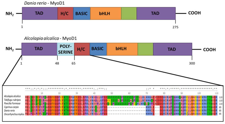
Figure 1
Inclusion of a polyserine insertion in MyoD1 in some fish species. Comparison of the protein structure of MyoD1 from Danio rerio (above) and Oreochromis (Alcolapia) alcalica (below). The enlargement of the schematic shows an alignment of the amino acid sequences from the indicated part of MyoD1 proteins from Oreochromis (Alcolapia) alcalica, Takifugu ruberipes, Poecilia formosa, Cyprinus carpio, Danio rerio, Oncorhynchus mykiss including the polyserine insertion in some species. Multiple species alignment of MyoD1 with and without the polyserine insertion. * represents exact match and: represents residues where the majority of species show conservation.