Fig. 3
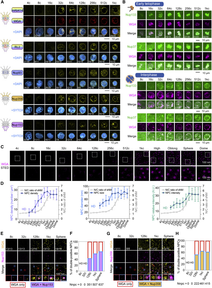
Figure 3. Gradual recruitment of NPC components during early embryogenesis (A) Detection of specific Nups in the nuclear rim in early embryos. DAPI or SYTOX, nuclei. The fraction of nuclei with the representative pattern was indicated at the bottom. Intranuclear signals could be residual NE embedded with NPCs. (B) NE/NPCs localization of transgenic Nup133-GFP and GFP-Nup37 proteins at indicated cell-cycle phases and stages. 100 pg/embryo of WGA-Alex647 was injected at 1c. (C and D) STED microscopy of individual NPCs with WGA-Alexa647 staining. Relative intensity was NPC intensity minus background intensity (same applied to following figures). HD, hardly detected. N/C ratio of sNM was recapitulated from Figure 2H. (E–H) Co-detection of Nup153 (E) or Nup358 (G) with WGA by STED microscopy. The boxed areas in the 3rd row were enlarged in the 4th row by color matching. The ratio of nuclei with representative NPC pattern was indicated in the top row. HD, hardly detected. Nnpc, NPC number. See also Figure S3 and Video S4.
Reprinted from Cell, 185(26), Shen, W., Gong, B., Xing, C., Zhang, L., Sun, J., Chen, Y., Yang, C., Yan, L., Chen, L., Yao, L., Li, G., Deng, H., Wu, X., Meng, A., Comprehensive maturity of nuclear pore complexes regulates zygotic genome activation, 4954-4970.e20, Copyright (2022) with permission from Elsevier. Full text @ Cell

