IMAGE

Fig. 6

ID
ZDB-IMAGE-230522-17
Source
Figures for Shin et al., 2023
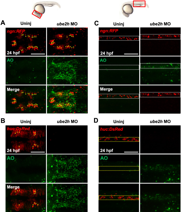
Image
Figure Caption

Fig. 6

Depletion of ube2h disrupts the maintenance of neurons during zebrafish embryogenesis. A, B Dorsal view of confocal microscopy images of AO staining (Green) of the brains of ube2h morphants and uninjected controls with the transgenic background of either ngn:RFP (A) or huc:DsRed (B). at 24 hpf. Yellow arrowheads indicate either ngn- or huc-positive cells (red). C, D Lateral view of confocal microscopy images of AO staining in the body trunk of ube2h morphants and uninjected controls with the ngn:RFP (C) and huc:DsRed (D) transgenic background. The white dotted box indicates the ngn-positive dorsal area, while yellow bracket indicates the huc-positive ventral area in the neural tube

Acknowledgments
This image is the copyrighted work of the attributed author or publisher, and ZFIN has permission only to display this image to its users. Additional permissions should be obtained from the applicable author or publisher of the image. Full text @ Hum. Genomics