
|
Fig. 5.
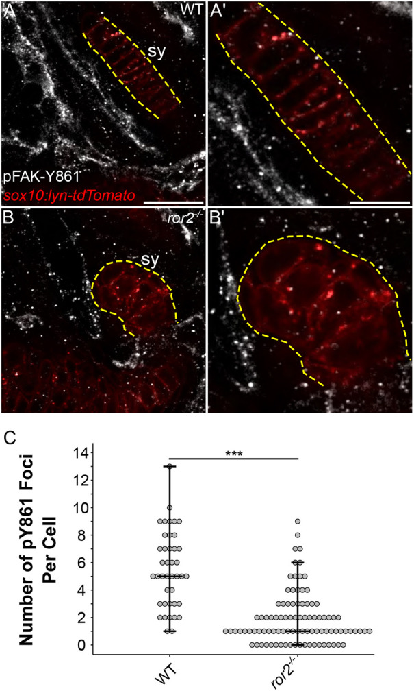
ror2 chondrocytes have a reduced number of focal adhesions. (A-B′) pFAK staining. Representative symplectic cartilages in WT (A) and ror2−/− (B) sox10:lyn-tdTomato transgenic fish at 3 dpf stained with anti-phospho-tyrosine 861 focal adhesion kinase antibody (pFAK-Y861) in white. Magnified views of symplectic cartilages in A′ and B′, respectively. Sox10:lyn-tdTomato chondrocytes in red. Panels A and B are a single slice of a z-stack. Dashed yellow lines delineate the symplectic cartilage visible in the selected slice. (C) Quantification of the number of pY861 foci per cell. Each point represents a single cell. Maximum, minimum and median values are depicted as crossbars on the dot plot. Sy, symplectic cartilage. ***P<0.001 (Wilcoxon rank-sum test). Scale bars: 20 μm for A and B; 10 μm for A′ and B′.