
|
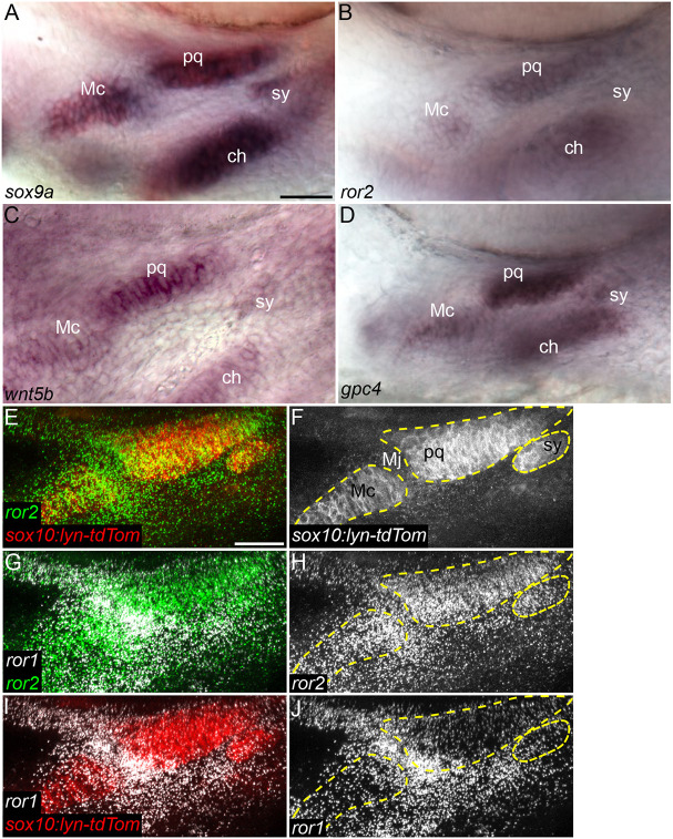
Fig. 1.
Wnt cell polarity pathway genes are expressed in cartilage progenitors. (A-D) In situ hybridization for sox9a (A), ror2 (B), wnt5b (C) and gpc4 (D) in 54 hpf wild-type (WT) embryos. Ventrolateral views of the mandibular and hyoid arches below the eye. (E-J) HCR for ror1 and ror2 in a 55 hpf WT Tg(sox10-lyn-tdTomato) (sox10:lyn-tdTom) embryo. ror1 in white, ror2 in green and sox10:lyn-tdTom in red (E,G,I). Grayscale (F,H,J). Panels E-J are z-projections. ch, ceratohyal cartilage; Mc, Meckel's cartilage; Mj, Meckel's joint; pq, palatoquadrate cartilage; sy, symplectic cartilage. Mc, pq and sy cartilages are outlined in dashed yellow line. Anterior to the left in all panels. Scale bars: 25 μm.