
|
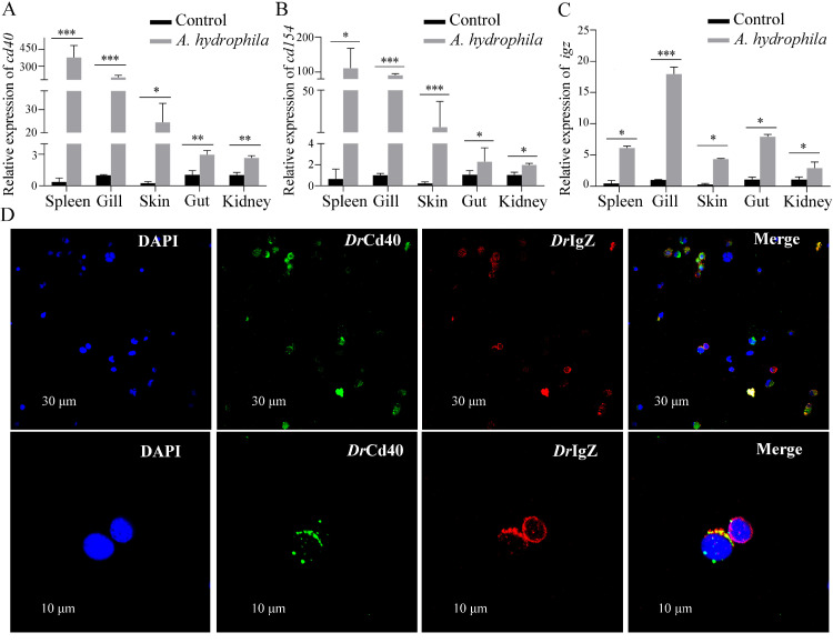
Fig. 1
Examination on the expression of cd40/cd154 and igz genes in immune-related tissues and distribution of Cd40 on IgZ+ B cells of zebrafish. (A − C) Real-time PCR analysis for the expression of Cd40, Cd154 and igz in spleen, gill, skin, gut and head kidney tissues in A. hydrophila-stimulated groups. All data were obtained from at least three independent experiments (n = 30). ⁎p < 0.05, ⁎⁎p< 0.01, ⁎⁎⁎p < 0.001. (D) Immunofluorescence staining of Cd40 and IgZ on leucocytes. Cells were stained with rabbit anti-Cd40 (green) together with mouse anti-IgZ (red). DAPI (blue) shows the location of the nuclei. The images were obtained using a two-photon laser scanning confocal microscope (Zeiss LSM-710; original magnification, 630×). Scale bars, 30 μm or 10 μm.