
|
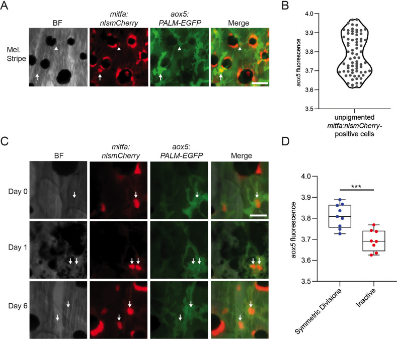
Figure 7
(A) Representative image of progenitors expressing different levels of aox5 promoter-driven PALM-EGFP. mitfa+aox5hi (arrowhead), mitfa+aox5lo (arrow). Scale bar = 50 µm. (B) Quantification of PALM-EGFP fluorescence intensity indicates groups of progenitors that express lower and higher levels of aox5. Mean pixel intensity per area; intensity values log normalized. n = 73 cells from 5 animals. (C) Images of an mitfa+aox5hi cell that underwent mitosis following melanocyte destruction. Scale bar = 30 µm. (D) Comparison of aox5 intensity in cells that underwent symmetric divisions or remained inactive. Mean pixel intensity per area; intensity values log normalized. n = 17 cells from 5 animals. p values calculated by Student’s t-test, ***p < 0.001.
aox5 expression predicts in vivo progenitor cell fate.