
|
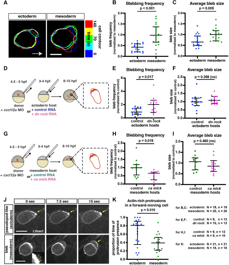
Fig. 2.
Alterations in protrusive behavior in PGCs located within different environments. (A) Overlay of cell contours over time of representative PGCs migrating in ectodermal and mesodermal tissues. Contours were aligned to the back of the cell to correct for forward movement. Arrow indicates the direction of migration. Scale bar: 10 µm. (B,C) Blebbing frequency (B) and average bleb size (C) of PGCs moving within converted embryos. For bleb frequency comparison (B), a Mann–Whitney test was performed; for average bleb size (C), a two-tailed t-test was performed. (D) The PGC transplantation experiment scheme. Labeled wild-type PGCs were transplanted into ectoderm-converted embryos that were injected at the one-cell stage with either control RNA or a dominant-negative form of ROCK. 4 to 6 h later, blebs formed by the transplanted PGCs were analyzed. (G) A similar transplantation was performed into mesoderm-converted embryos that expressed either control RNA or RNA encoding a constitutively active form of MLCK. (E,F,H,I) Bleb formation frequency (E,H) and average bleb size (F,I) produced by PGCs transplanted into the different environments. Two-tailed t-tests were performed. (J) Snapshots of migrating PGCs. Arrows indicate actin enrichment at the edge of protrusion. Inset shows a zoomed-in, contrast-adjusted view of the early-stage bleb, with a yellow dashed line outlining the bleb contour. Scale bars: 15 µm. (K) The proportion of time actin-rich protrusions that are present at the front of a forward-moving cell. Mann–Whitney test was used. The graphs show the median; whiskers indicate the interquartile range (IQR). N and n at the bottom right represent numbers of embryos and cells, respectively.