
|
Fig. 2.
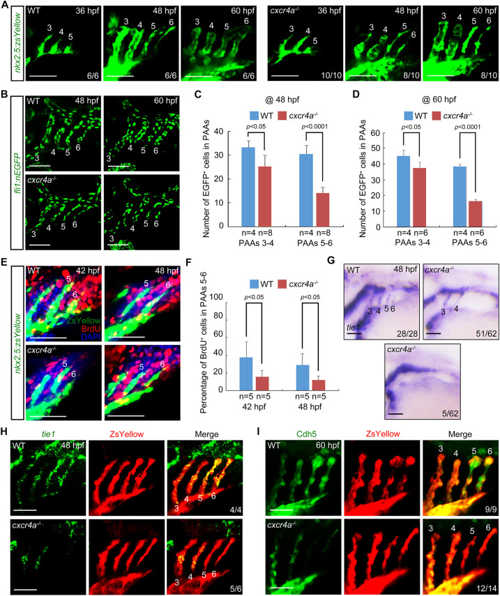
Lack of cxcr4a represses both the proliferation and differentiation of PAA angioblasts. (A) Time-lapse recording of ZsYellow fluorescence in the pharyngeal regions of wild-type (WT) and cxcr4a−/− embryos in Tg(nkx2.5:ZsYellow) background. (B-D) cxcr4a−/− mutants showed a significantly reduced cell number in posterior PAAs. Representative confocal sections of the PAAs in wild-type (WT) control and cxcr4a-deficient embryos are shown in B. Quantified cell numbers in PAAs 3-6 are shown in C and D. Error bars indicate s.d. of three biological replicates. (E,F) Detection of proliferative PAA angioblasts. Wild-type (WT) and cxcr4a−/− embryos were co-immunostained using anti-ZsYellow (green) and anti-BrdU (red) antibodies. Nuclear DNA was stained using DAPI (blue). Representative pictures are shown in E and the percentage of BrdU-positive cells in PAAs 5 and 6 for each group is shown in F. Error bars indicate s.d. of three biological replicates. (G,H) Expression analysis of tie1 in the wild-type (WT) and cxcr4a−/− embryos at 48 hpf by in situ hybridization (G) and fluorescent in situ hybridization combined with immunostaining (H). (I) Cdh5 levels were greatly decreased in the posterior PAAs of cxcr4a−/− mutants. The indicated embryos were immunostained using Cdh5 (green) and ZsYellow (red) antibodies. Scale bars: 50 μm.