
|
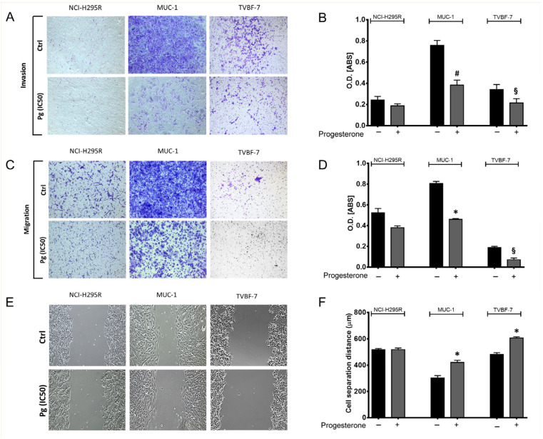
Figure 3
Pg suppressed ACC cell invasion (A) and migration (C) ability in transwell assays. Images were acquired using an Olympus IX51 optical microscope at 10× magnification. (B,D) Quantification of the number of invasive and migrated cells was analyzed using the absorbance of the staining detected at 560 nm. (E) Representative images of wound healing assay used to detect migrated ACC cells. (F) The distance between each edge of the scratch was measured using NIH ImageJ Software V 1.52a. Images were acquired using an Olympus IX51 optical microscope equipped with a 10× objective. Data are shown as the mean ± SEM of three independent experiments. * p < 0.0001, # p < 0.001, § p < 0.05 vs. untreated cells.