
|
Figure 5
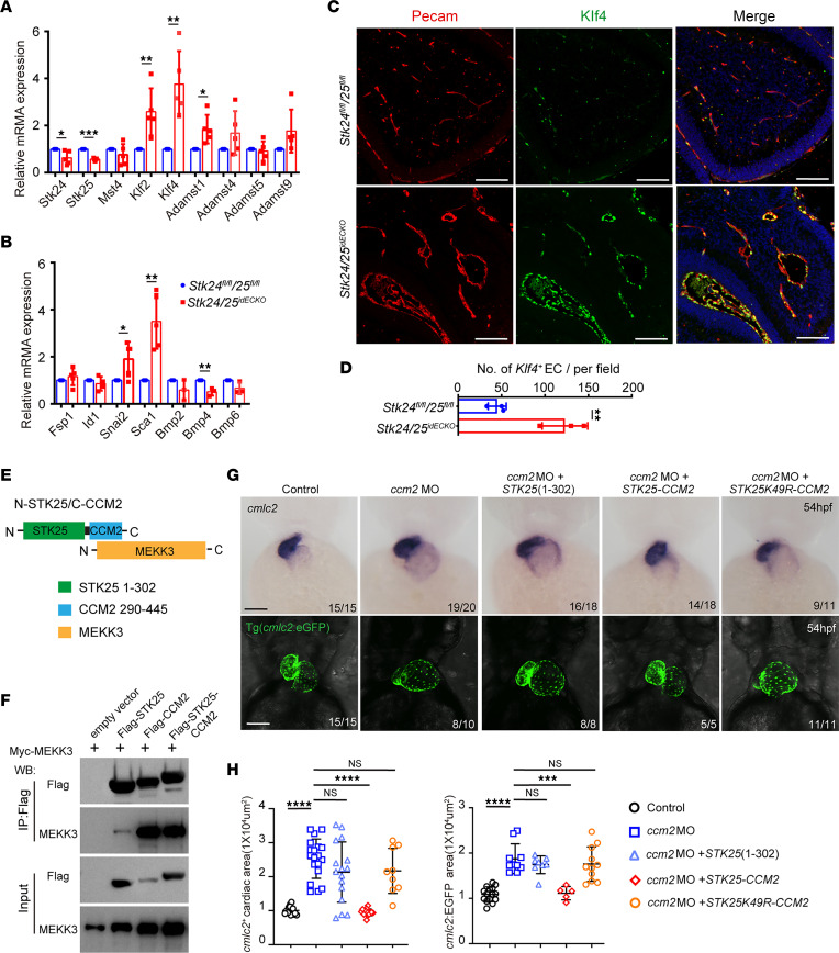
(A and B) Relative mRNA expression level of CCM-related genes in endothelial cells isolated from control and Stk24/25idECKO mice at P6 after induction at P2. n = 5 for each group — except Bmp2, Bmp4, and Bmp6, for which n = 3 was used. (C) Pecam and Klf4 immunofluorescence staining in endothelial cells of Stk24fl/fl;Stk25fl/fl (n = 3) and Stk24/25idECKO mouse brains (n = 3) at P6 after induction at P2. Scale bars: 100 μm. (D) Quantitative analysis showing increased Klf4+ EC in Stk24/25idECKO mice compared with Stk24fl/fl;Stk25fl/fl mice. The quantitative data (mean ± SD) from 3 independent experiments are reported, and significance was determined using unpaired t test. **P < 0.01. (E) Schematic representation of the interaction between MEKK3 and STK25-CCM2 hybrid protein consists of N terminal kinase domain of STK and C terminal MEKK3 interacting domain of CCM2. (F) Immunoprecipitation experiment shows that STK25/CCM2 hybrid protein interaction with MEKK3 was comparable with that of CCM2. (G) Representative images of in situ staining of cmlc2 and fluorescence imaging of the hearts of Tg (cmlc2:EGFP) zebrafish embryos in which myocardial cells express EGFP. The ccm2 morpholino induced dilated heart, while coinjection with mRNA expressing STK25-CCM2 rescued the dilated heart phenotype compared with injection of mRNA only expressing STK25(1-302) or STK25K49R-CCM2 hybrid protein. Scale bars: 100 μm. (H) Quantification of cmlc2+ cardiac area and cmlc2:EGFP area of zebrafish embryos with ccm2 morpholino and different cRNA. Data are presented as mean ± SD, and significance was determined using unpaired t test (A, B, and D) or 1-way ANOVA (H). *P < 0.05, **P < 0.01, ***P < 0.001, ****P < 0.0001.