
Fig. 1
- ID
- ZDB-IMAGE-230403-23
- Genes
- Publication
- Heidary et al., 2023 - A zebrafish model of growth hormone insensitivity syndrome with immune dysregulation 1 (GHISID1)
- All Figures
- Figures for Heidary et al., 2023
|
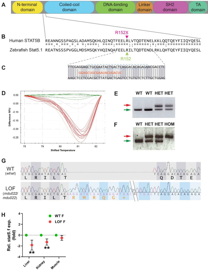
Fig. 1
Generation of a zebrafish Stat5.1 mutation mimicking those in human STAT5B associated with GHISID1. A Schematic diagram of the STAT5B protein domain architecture consisting of N-terminal (yellow), coiled-coil (blue), DNA-binding (green), linker (orange), SH2 (pink), and transactivation (TA) (dark green) domains. B Alignment of human STAT5B with zebrafish stat5.1 sequences around the site of the R152X mutation (green) seen in human GHISID1, showing identical (*), highly similar (:) and similar (.) amino acids. C Nucleotide sequence of zebrafish stat5.1 genomic DNA targeted using CRISPR/Cas9 with the gRNA (orange) shown and the coding phase indicated. D HRM analysis of a sample of embryos injected with stat5.1 gRNA and Cas9, with wild-type embryos in green and other colours representing potential mutants.