
|
Figure 1
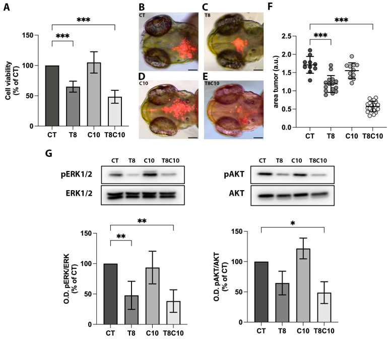
Hh and HDAC6 inhibition impaired U87-MG cell viability. (A) MTT assay of U87-MG cells treated for 48 h with vehicle (CT), 8 μM of TubA (T8), 10 μM of cyclo (C10) and 8 μM of TubA + 10 μM cyclo (T8C10). (B–E) Representative images of the head region of 3 dpf zebrafish embryos xenotransplanted with labeled U87-MG cells pretreated with (B) CT, (C) T8, (D) C10, and (E) T8C10. (F) Quantification of the tumor area at t1 (24 h postinjection, hpi) normalized to the tumor area at t0 (immediately after U87 injection). (G) Western blot analyses and quantification of pERK and pAKT protein expression levels following 48 h treatments with or without T8 and C10, alone or in combination. Data are expressed as the percentage of the control (A,G) or mean ± standard deviation (F). CT—vehicle; TubA/T—tubastatin A; cyclo/C—cyclopamine. One-way ANOVA with Tukey post hoc correction. *** p < 0.001; ** p < 0.01; * p < 0.05; nonsignificant data are not shown. Scale bare indicates 100 μm.