
Figure 3
- ID
- ZDB-IMAGE-230331-68
- Publication
- Yuan et al., 2023 - Roles of miR-196a and miR-196b in Zebrafish Motor Function
- All Figures
- Figures for Yuan et al., 2023
|
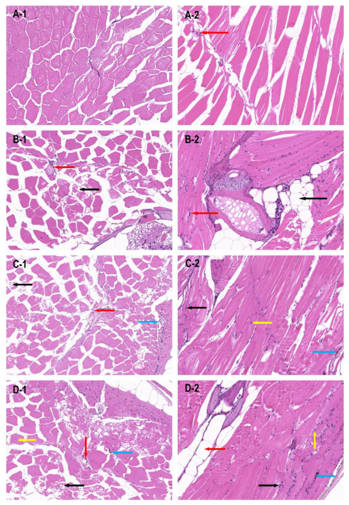
Figure 3
HE staining results of dorsal muscle tissue of zebrafish in different groups (20×).The dorsal muscle tissue was damaged when the miR-196a-1 and miR-196b genes were knocked out. (A) wild-type zebrafish; (B): zebrafish with miR-196a-1 gene knockout; (C): miR-196b gene knockout zebrafish; (D): zebrafish with miR-196a-1 and miR-196b gene knockout; -1 is the transverse section of muscle tissue, -2 is the longitudinal section of muscle tissue. The red arrow points to inflammatory cell infiltration, the black arrow points to vacuolar degeneration of muscle fibers, the blue arrow points to melanin deposition, the yellow arrow points to inward nuclear movement.