Image
Figure Caption
Fig. 2
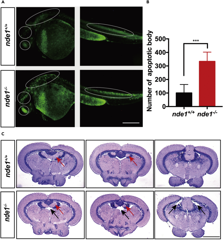
Morphological analysis and apoptosis analysis of nde1−/−
(A) AO staining of nde1+/+ and nde1−/− at 30hpf; white dotted oval indicates telencephalon, midbrain, rhombomere, and spine, respectively. Scale bar: 0.2 mm.
(B) Statistics of apoptotic bodies after AO staining in nde1+/+ and nde1−/−.
(C) HE-stained paraffin sections of midbrain of nde1+/+ and nde1−/−. Black arrow indicates the gaps between the ventricles; red arrow indicates valvula cerebelli. Scale bar: 0.5 mm. ∗p < 0.05, ∗∗p < 0.01, ∗∗∗p < 0.001. Data are represented as mean ± SEM
Figure Data
Acknowledgments
This image is the copyrighted work of the attributed author or publisher, and
ZFIN has permission only to display this image to its users.
Additional permissions should be obtained from the applicable author or publisher of the image.
Full text @ iScience

