
Fig 9
- ID
- ZDB-IMAGE-230315-59
- Publication
- Xie et al., 2023 - Ependymal polarity defects coupled with disorganized ciliary beating drive abnormal cerebrospinal fluid flow and spine curvature in zebrafish
- All Figures
- Figures for Xie et al., 2023
|
Fig 9
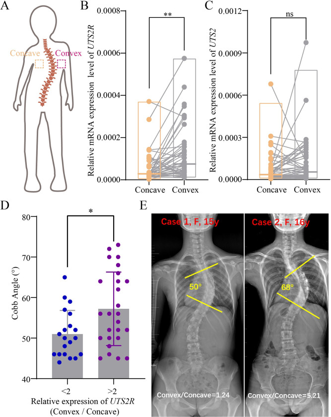
(A) Diagram indicating the position of bilateral paravertebral muscles harvested from AIS patients. (B, C) Relative expression of UTS2R and UTS2 in bilateral paravertebral muscles of AIS patients (n = 46). (D) Distribution of Cobb angles within the two groups of AIS patients exhibiting a ratio of UTS2R expression on the convex versus concave sides greater (>) or less (<) than 2. (E) Representative X-ray images showing two AIS patients with convex/concave ratios greater (5.21) or less than 2 (1.24). Measurements of Cobb angles are also indicated. The data underlying the graphs shown in the figure can be found in S1 Data.