
|
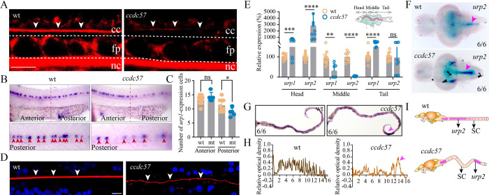
Fig 7
(A) Representative images of Reissner fiber (RF, white arrows) in 2 dpf wild type and ccdc57 mutant larvae. cc, central canal; nc, notochord; fp, floor plate. (B) Whole-mount in situ hybridization results showing the expression of urp1 in 24 hpf control and ccdc57 mutant larvae as indicated. The enlarged views of the staining in the posterior region are shown in the bottom. (C) Statistical analysis showing the number of urp1-expressing cells in the anterior and posterior part of the trunk. (D) Confocal images showing RF (white arrow) in wild type and ccdc57 adult mutants. RF was stained with wheat germ agglutinin (WGA, red), and nuclei were counterstained with DAPI. (E) qPCR analysis showing the expression of urotensin genes (urp1 and urp2) in different parts of the adult trunk as illustrated in the diagram. (F) In situ hybridization results showing the expression of urp2 in the brains of adult wild type and ccdc57 mutant as indicated. Arrows point to the expression of urp2 in the posterior part of the brain. (G) In situ hybridization results showing the expression of urp2 in the spinal cord of wild type and ccdc57 mutant as indicated. The purple arrowhead indicates the sites of enriched urp2 expression. (H) The line graphs showing the relative optical density of the expression of urp2 in wild type and ccdc57 mutants. (I) Model illustrating the distribution of urp2 in wild type and ccdc57 mutant. Scale bars: 7.5 μm in panel A; 10 μm in panel D. The data underlying the graphs shown in the figure can be found in S1 Data.