
|
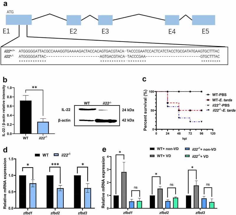
Figure 2.
IL-22 mediated VD-induced β-defensin expression in zebrafish intestine. (a) The deletion site by CRISPR/Cas9 on the il22 gene exon (E)1 (exons are in blue boxes) was displayed. (b) The protein level of IL-22 in the intestine of WT and il22-/- zebrafish was compared (n = 6/group). The image is representative of 6 replicates. (c) Zebrafish at 3 mpf were i.p. injected with 107 CFU E. tarda or PBS, and the survival rate was recorded until 96 hours-post infection (n = 10/group). (d) The gene expression of zfbd1, zfbd2 and zfbd3 in zebrafish intestine was measured. (e) After WT and il22 mutant zebrafish at 2 mpf were fed with 0 or 800 IU/kg dietary VD3 for 4 weeks, the transcript levels of zfbd1, zfbd2 and zfbd3 in zebrafish intestine were evaluated (n = 6–8/group). *p < 0.05, ***p < 0.001, ns: non-significance. See also Figures S2.