
|
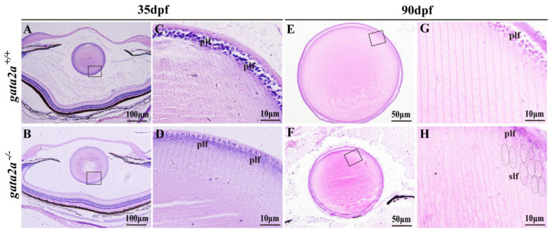
Figure 4
Histological analysis of the eyes from WT and gata2a mutants at 35 and 90 dpf. (A–D) Histology of the eyes at 35 dpf, the size of the lens from gata2a mutants were similar to that of WT (A,B), while most of the primary lens fiber cells were degenerated (C,D). (E–H) Lens from gata2a mutants were smaller and opaque compared to those from WT siblings (E,F). Most of the primary lens fiber cells in the gata2a mutants were degenerated, and the nuclei were not degraded in some of the secondary lens fiber cells (G,H). (C,D,G,H) are magnifications of the boxed areas in (A,B,G,H), respectively. dpf: day post fertilization. plf: primary lens fiber cell; slf: secondary lens fiber cell. The dotted circles and ellipses in the (H) diagram represent primary and secondary lens fiber cells, respectively.