
|
Fig. 4.
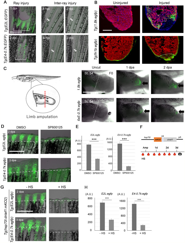
Conservation of E-box/AP-1-mediated regeneration response beyond tissue types and species. (A) EGFP induction by ray injury, but not by inter-ray incision in Tg(E2L:egfp) (upper) and Tg(E4-0.7k:egfp) (lower). EGFP induction was prominently detected on the proximal side of the injury. Arrow, site of inter-ray injury. Scale bar: 500 µm. (B) EGFP induction after heart resection in Tg(1.8k:egfp) (upper) and Tg(fn1b:egfp) (lower). MF20, cardiac muscles. Tg(1.8k:egfp) showed EGFP induction in the cardiomyocytes around amputation (n=5 for uninjured and injured fish). In Tg(fn1b:EGFP), EGFP expression in the epicardial cells can be observed in the uncut heart (n=3 fish), but it was upregulated by heart resection (n=3 fish). Dotted line, amputation site. Scale bar: 200 µm. (C) EGFP induction in the amputated limb bud of X. laevis injected with respective constructs at the one-cell stage. Arrows, EGFP expression; dotted lines, site of amputation. EGFP induction was observed in mesenchymal cells (1.8k:egfp, n=12 of 12 with lens EGFP+ tadpoles) and the distal tip of epithelial cells (6xE-0.7k:egfp, n=8 of 8 with lens EGFP+ tadpole). Scale bar: 1 mm. (D) Knockdown of the EGFP induction by the JNK inhibitor, SP600125, in Tg(E2L:egfp) (n =10) and Tg(E4-0.7k:egfp) (n =10). Dotted lines, amputation plane; brackets, cells forming the WE (upper panel) and blastema (lower panel). Scale bar: 0.5 mm. (E) Quantification of EGFP fluorescence in (D). Data are presented as mean ±s.e.m. Student's t-test. ***P<0.001. (F) dnLef1 construct and the experimental procedure. (G) Knockdown of EGFP induction by dnLef1 overexpression in Tg(E2L:egfp) (n=5) and Tg(E4-0.7k:egf) (n=12). Dotted lines, amputation plane; brackets, cells forming the WE (upper panel) and blastema (lower panel). Scale bar: 0.5 mm. (H) Quantification of EGFP fluorescence in G. Data are presented as mean ±s.e.m. Student's t-test. ***P<0.001.