
Image
|
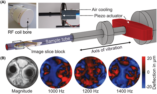
Figure Caption
Fig. 1 Setup for MRE of zebrafish in a 7T preclinical MRI scanner. (A) A piezoelectric actuation unit, shown in red, generates harmonic oscillations synchronized to the MRE sequence. The principal vibration direction is along the cylinder axis, which results in predominantly cylindrical waves. The box demarcates the area covered by the imaging slice slab. The custom volume resonator coil is also shown. (B) MRE magnitude image (grayscale) and wave images of different frequencies (color scale) showing the through-plane wave component, which was encoded by the sequence. MRE, MR elastography; T, tesla
Acknowledgments
This image is the copyrighted work of the attributed author or publisher, and
ZFIN has permission only to display this image to its users.
Additional permissions should be obtained from the applicable author or publisher of the image.
Full text @ Magn Reson Med