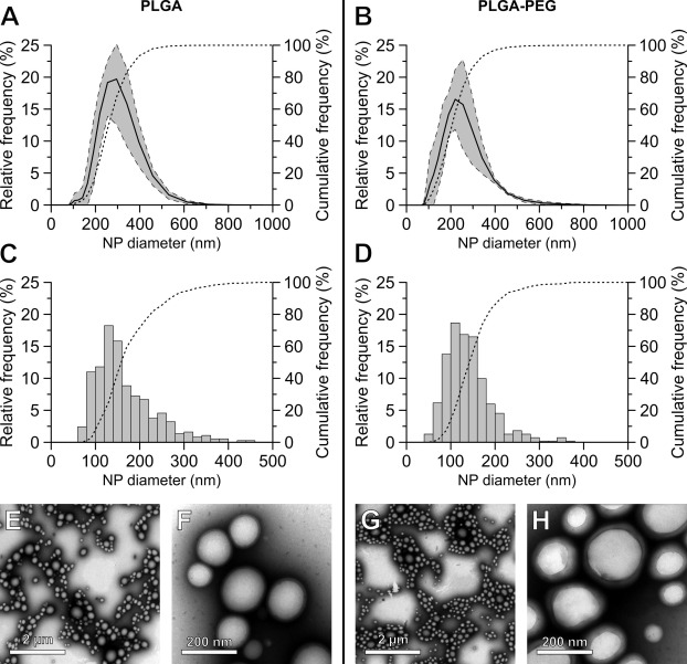
Fig. 3 Size distribution and morphology of CPZ-loaded PLGA and PLGA-PEG nanoparticles. (A,B): DLS measurements for the diameter of CPZ-loaded PLGA (A) and PLGA-PEG (B) nanoparticles. Each graph shows the average count-based distribution in size (d.nm) of three separate nanoparticle batches, analysed by DLS within 3 h of production. The dashed lines indicate the standard deviation and the dotted line represent the cumulative distribution. (C-H): Freshly prepared CPZ-loaded nanocarriers were stained with uranyl acetate and imaged with TEM. (C,D): Analysis of TEM images for the size distribution (d.nm) of nanoparticles made with PLGA (C) and PLGA-PEG (D). The dotted lines indicate the cumulative distribution frequency. TEM images are shown of CPZ-loaded PLGA (E,F) and PLGA-PEG (G,H). Scale bars represent either 2 µm (E,G) or 200 nm (F,H).
Image
Figure Caption
Acknowledgments
This image is the copyrighted work of the attributed author or publisher, and
ZFIN has permission only to display this image to its users.
Additional permissions should be obtained from the applicable author or publisher of the image.
Full text @ Int J Pharm

